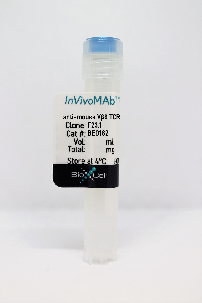
About InVivoMAb anti-mouse Vβ8 TCR
The F23.1 monoclonal antibody reacts with the Vβ 8.1, Vβ 8.2, and Vβ 8.3 TCR (V beta 8.1-3 T cell receptors) of mice having the b haplotype of the Tcrb gene complex (e.g., AKR, BALB/c, C3H, C57BL, DBA/1, DBA/2). The TCR is expressed on the surface of T lymphocytes and is responsible for recognizing fragments of antigen as peptides bound to MHC molecules. When the TCR engages with antigenic peptide and MHC the T lymphocyte is activated through signal transduction. The F23.1 antibody has been shown to activate Vβ 8 TCR-expressing T lymphocytes, as well as block cytolysis mediated by Vβ 8 TCR-expressing cytotoxic T lymphocytes. Additionally, in vivo treatment of neonatal mice with the F23.1 antibody can arrest intrathymic maturation of Vβ 8 TCR-expressing T cells.
InVivoMAb anti-mouse Vβ8 TCR Specifications
| Isotype | Mouse IgG2a, κ |
| Recommended Isotype Control(s) | InVivoMAb mouse IgG2a isotype control, unknown specificity |
| Recommended Dilution Buffer | InVivoPure pH 7.0 Dilution Buffer |
| Immunogen | BALB.B Mouse lymph node and spleen T cells |
| Reported Applications | Flow cytometry |
| Formulation | PBS, pH 7.0 Contains no stabilizers or preservatives |
| Endotoxin | <2EU/mg (<0.002EU/μg) Determined by LAL gel clotting assay |
| Purity | >95% Determined by SDS-PAGE |
| Sterility | 0.2 μm filtered |
| Production | Purified from cell culture supernatant in an animal-free facility |
| Purification | Protein G |
| RRID | AB_10950392 |
| Molecular Weight | 150 kDa |
| Storage | The antibody solution should be stored at the stock concentration at 4°C. Do not freeze. |
Application References
InVivoMAb anti-mouse Vβ8 TCR (CLONE: F23.1)
Bothur, E., et al (2015). "Antigen receptor-mediated depletion of FOXP3 in induced regulatory T-lymphocytes via PTPN2 and FOXO1" Nat Commun 6(8576): doi:10.1038/ncomms9576. PubMed
Regulatory T-cells induced via IL-2 and TGF in vitro (iTreg) suppress immune cells and are potential therapeutics during autoimmunity. However, several reports described their re-differentiation into pathogenic cells in vivo and loss of their key functional transcription factor (TF) FOXP3 after T-cell antigen receptor (TCR)-signalling in vitro. Here, we show that TCR-activation antagonizes two necessary TFs for foxp3 gene transcription, which are themselves regulated by phosphorylation. Although the tyrosine phosphatase PTPN2 is induced to restrain IL-2-mediated phosphorylation of the TF STAT5, expression of the TF FOXO1 is downregulated and miR-182, a suppressor of FOXO1 expression, is upregulated. TGF counteracts the FOXP3-depleting TCR-signal by reassuring FOXO1 expression and by re-licensing STAT5 phosphorylation. Overexpressed phosphorylation-independent active versions of FOXO1 and STAT5 or knockdown of PTPN2 restores FOXP3 expression despite TCR-signal and absence of TGF. This study suggests novel targets for stabilisation and less dangerous application of iTreg during devastating inflammation.
Rojo, J. M., et al (2014). "Characteristics of TCR/CD3 complex CD3{varepsilon} chains of regulatory CD4+ T (Treg) lymphocytes: role in Treg differentiation in vitro and impact on Treg in vivo" J Leukoc Biol 95(3): 441-450. PubMed
Tregs are anergic CD4(+)CD25(+)Foxp3(+) T lymphocytes exerting active suppression to control immune and autoimmune responses. However, the factors in TCR recognition underlying Treg differentiation are unclear. Based on our previous data, we hypothesized that Treg TCR/CD3 antigen receptor complexes might differ from those of CD4(+)CD25(-) Tconv. Expression levels of TCR/CD3, CD3epsilon,zeta chains, or other molecules involved in antigen signaling and the characteristics of CD3epsilon chains were analyzed in thymus or spleen Treg cells from normal mice. Tregs had quantitative and qualitatively distinct TCR/CD3 complexes and CD3epsilon chains. They expressed significantly lower levels of the TCR/CD3 antigen receptor, CD3epsilon chains, TCR-zeta chain, or the CD4 coreceptor than Tconv. Levels of kinases, adaptor molecules involved in TCR signaling, and early downstream activation pathways were also lower in Tregs than in Tconv. Furthermore, TCR/CD3 complexes in Tregs were enriched in CD3epsilon chains conserving their N-terminal, negatively charged amino acid residues; this trait is linked to a higher activation threshold. Transfection of mutant CD3epsilon chains lacking these residues inhibited the differentiation of mature CD4(+)Foxp3(-) T lymphocytes into CD4(+)Foxp3(+) Tregs, and differences in CD3epsilon chain recognition by antibodies could be used to enrich for Tregs in vivo. Our results show quantitative and qualitative differences in the TCR/CD3 complex, supporting the hyporesponsive phenotype of Tregs concerning TCR/CD3 signals. These differences might reconcile avidity and flexible threshold models of Treg differentiation and be used to implement therapeutic approaches involving Treg manipulation.
Yang, Y., et al (2014). "Focused specificity of intestinal TH17 cells towards commensal bacterial antigens" Nature 510(7503): 152-156. PubMed
T-helper-17 (TH17) cells have critical roles in mucosal defence and in autoimmune disease pathogenesis. They are most abundant in the small intestine lamina propria, where their presence requires colonization of mice with microbiota. Segmented filamentous bacteria (SFB) are sufficient to induce TH17 cells and to promote TH17-dependent autoimmune disease in animal models. However, the specificity of TH17 cells, the mechanism of their induction by distinct bacteria, and the means by which they foster tissue-specific inflammation remain unknown. Here we show that the T-cell antigen receptor (TCR) repertoire of intestinal TH17 cells in SFB-colonized mice has minimal overlap with that of other intestinal CD4(+) T cells and that most TH17 cells, but not other T cells, recognize antigens encoded by SFB. T cells with antigen receptors specific for SFB-encoded peptides differentiated into RORgammat-expressing TH17 cells, even if SFB-colonized mice also harboured a strong TH1 cell inducer, Listeria monocytogenes, in their intestine. The match of T-cell effector function with antigen specificity is thus determined by the type of bacteria that produce the antigen. These findings have significant implications for understanding how commensal microbiota contribute to organ-specific autoimmunity and for developing novel mucosal vaccines.
Zhu, L., et al (2013). "A transgenic TCR directs the development of IL-4+ and PLZF+ innate CD4 T cells" J Immunol 191(2): 737-744. PubMed
MHC class II-expressing thymocytes can efficiently mediate positive selection of CD4 T cells in mice. Thymocyte-selected CD4 (T-CD4) T cells have an innate-like phenotype similar to invariant NKT cells. To investigate the development and function of T-CD4 T cells in-depth, we cloned TCR genes from T-CD4 T cells and generated transgenic mice. Remarkably, positive selection of T-CD4 TCR transgenic (T3) thymocytes occurred more efficiently when MHC class II was expressed by thymocytes than by thymic epithelial cells. Similar to polyclonal T-CD4 T cells and also invariant NKT cells, T3 CD4 T cell development is controlled by signaling lymphocyte activation molecule/signaling lymphocyte activation molecule-associated protein signaling, and the cells expressed both IL-4 and promyelocytic leukemia zinc finger (PLZF). Surprisingly, the selected T3 CD4 T cells were heterogeneous in that only half expressed IL-4 and only half expressed PLZF. IL-4- and PLZF-expressing cells were first found at the double-positive cell stage. Thus, the expression of IL-4 and PLZF seems to be determined by an unidentified event that occurs postselection and is not solely dependent on TCR specificity or the selection process, per se. Taken together, our data show for the first time, to our knowledge, that the TCR specificity regulates but does not determine the development of innate CD4 T cells by thymocytes.

